Dental x-rays are necessary for anyone who wants to maintain a healthy mouth. X-rays allow your dentist to look through the tooth, thus being able to diagnose cavities, gum disease, and developing abscesses in their early stages.
Without dental x-rays, you cannot know for certain if your teeth and jawbone are truly healthy.
Let’s begin with the question: Are dental x-rays actually necessary at all? It is true that there are some conditions your dentist can see and diagnose with the naked eye. Problems like large cavities, receding gums, or loose teeth are often evident without the assistance of x-rays. Some infections are also visible, while some remain hidden within the jawbone.
The need for dental x-rays lies in the ability and responsibility of your dentist to detect problems as early as possible. Dental issues like cavities and gum disease require treatments that are less extensive and less expensive the earlier you catch them. When someone waits until a problem causes noticeable symptoms (like a toothache) or visible evidence (like a large, black hole in the tooth), the problem is already into its advanced stages.
Dental x-rays are necessary for anyone who wants to maintain a healthy mouth through prevention and early detection. X-rays allow your dentist to look through the tooth at each different layer, thus being able to diagnose cavities, gum disease, and developing abscesses in their initial stages.
Without dental x-rays, you cannot know for certain whether your teeth and jawbone are truly healthy.
Dental x-rays give your dentist an inside picture of what is going on under the surface of the teeth. X-Rays can also show your bone and the supporting structures of your mouth covered by gum tissue. Most dental diseases are progressive in nature, meaning they will get worse over time if left untreated. With that in mind, some of the diseases and disorders listed here will likely make their presence known eventually, but that often will only be after noteworthy damage has already occurred.
Cavities Between Teeth
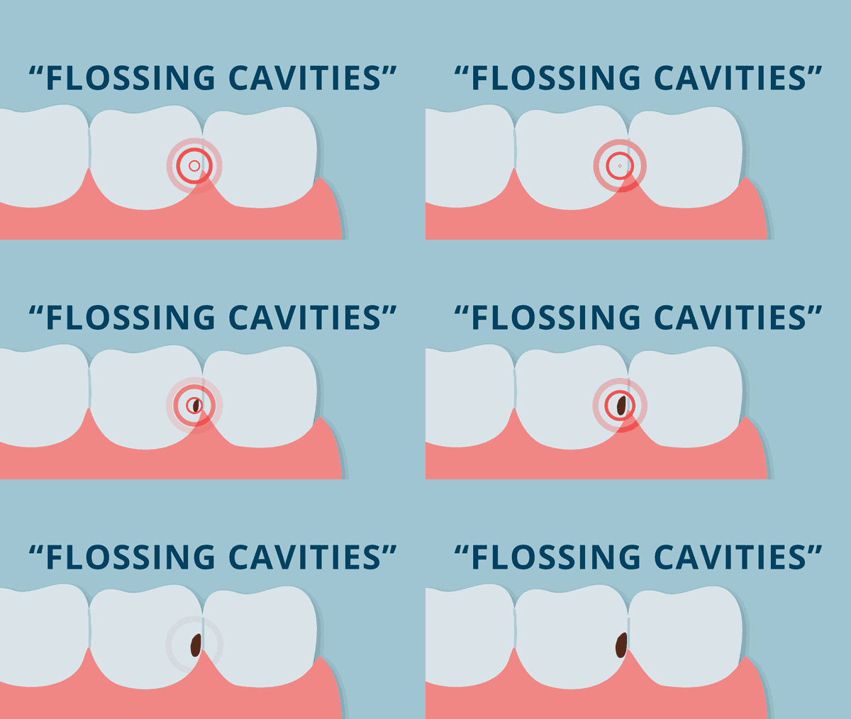
Many cavities develop in between your teeth where you floss. Plaque collects between the teeth, and if not removed consistently and effectively, it weakens tooth enamel and leads to cavities.

These “flossing cavities” begin on the side of the tooth and work their way inward, toward the center of the tooth. This type of cavity only becomes visible to the naked eye once it destroys enough hard tooth structure to form an actual hole in the enamel. Many cavities of this kind cannot be detected without an x-ray. Dental x-rays allow your dentist to use minimally invasive techniques to treat the progression of decay prior to needing more complex (and more expensive) dental procedures. This is why early detection with x-rays is vital for our oral health!
Unerupted, Impacted and Missing Teeth
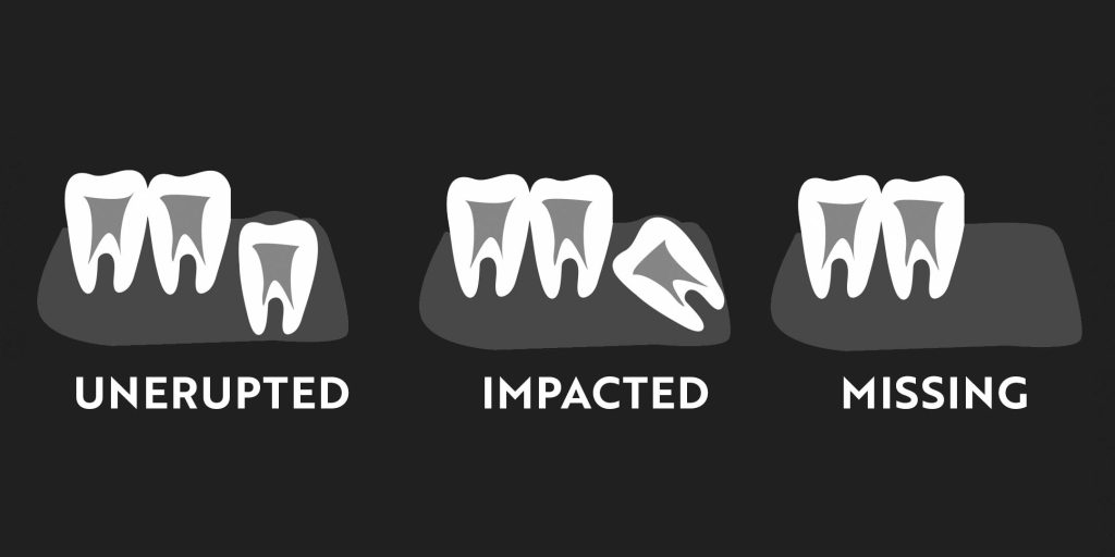
Dental x-rays are useful in finding teeth that do not appear inside the mouth when they should. When someone is “missing” a tooth at the time it should be present, it is important to determine whether that tooth is present and simply behind schedule, present but unable to erupt, or completely absent from the jaw.

An unerupted tooth exists in the jawbone in its correct position and orientation. It simply has not yet made its way into the dental arch.
An impacted tooth is present in the jawbone, but it is NOT in the correct position or orientation. Impacted teeth cannot erupt into the dental arch without dental intervention. This is a common scenario with wisdom teeth. A dental x-ray is necessary for proper diagnosis and treatment planning in the removal of impacted teeth.
A missing tooth is one that was never developed in the jaw. We refer to this situation as a “congenitally missing tooth.” A missing tooth or teeth will have an impact on a person’s smile. The patient can use orthodontic treatment to align the surrounding teeth properly, or the patient can choose to replace the missing tooth with a prosthetic tooth.
Tumors
There are a variety of tumors that can arise in the jawbone. These tumors develop from abnormal cells around the teeth or from the bone itself. Most of these lesions are benign and do not spread to other sites in the body as oral cancer can. However, they can grow, and their growth can cause expansion of the jawbone and movement of the nearby teeth. Consistent x-rays can lead to early detection of tumors. Once a tumor has grown substantially, a patient will likely need major intervention like surgery.
Jaw Fractures
Dental x-rays detect fractures in the jawbone that result from trauma. When someone has an accident that causes injury to the face, emergency issues like blood loss or pain are always addressed first. Other symptoms like swelling, headaches, or a change in your bite may not be noticed right away, but can often be attributed to a jaw fracture, best diagnosed by a dental x-ray.
Degenerative Changes in the Jaw Joints
The jaw joints, like any joint in the body, can undergo changes due to arthritis or degenerative joint disease. Degenerative changes can lead to a shift in the way the teeth bite together and significant TMJ symptoms.
Not all dental x-rays are alike. Different x-rays capture images of different areas of the upper and lower jaws, and they produce different levels of detail. Your dentist uses a variety of dental x-rays to capture a comprehensive view of your teeth and jawbone.
We can classify dental x-rays as either intraoral or extraoral. Intraoral x-rays are those that capture the image on film or a sensor inside the mouth. These are usually close-up views of the teeth in high definition. Extraoral x-rays capture an image of a much larger portion of the head on a film or sensor outside the mouth. This type of image provides a more broad view of the upper and lower jaws and is not as detailed as intraoral images.
Bitewing X-rays
Most dental patients have bitewing x-rays on a yearly basis at minimum. These small intraoral x-rays show a close-up view of three to four upper and lower teeth biting together. Bitewings are commonly used to show between-teeth cavities or tartar build up. They also show a small amount of the surrounding jawbone and can help identify signs of bone loss due to periodontal disease.
Periapical X-rays
Periapical x-rays are also close-up intraoral images, like bitewings. Rather than simultaneously showing upper and lower teeth, they show the full length of teeth on either the upper or lower. These x-rays are essential for monitoring the health of the roots of the teeth and when monitoring any root canal treatments and dental implants present in the mouth.
Occlusal X-rays
Typically used for children only, these small images capture the baby teeth and underlying permanent teeth. Taken only in the front of the mouth, occlusal images are more comfortable than other intraoral x-rays for small children. Dentists use occlusal x-rays to identify growth patterns and the health of developing permanent teeth.
Panoramic X-ray
A panoramic x-ray is a large extraoral scan that captures the entirety of the upper and lower jaws as a singular image. This type of x-ray machine rotates around the head while the patient bites onto a mouthpiece to hold the jaws in a precise location. This x-ray is beneficial in allowing your dentist to rule out any tumors or TMJ issues.
CBCT
CBCT stands for Cone Beam Computed Tomography, and is a relatively new form of dental imaging. The CBCT machine looks similar to a panoramic x-ray machine, but provides a three-dimensional image. With this 3D image, your dentist is able to gather more detailed information about the teeth and jaws. This is exceptionally valuable in planning the extraction of teeth, root canal treatments, and dental implant placements.
Yes!

All x-rays require a level of radiation to penetrate the human body and produce the desired image. The word radiation can sometimes cause fear, because we know overexposure can lead to serious long-term health defects like cancer. But it’s important to understand radiation is actually present all around us every day of our lives. Radiation occurs naturally in the air we breath, ground we walk on, and even in the foods we eat.
While there are some individuals with a higher risk for tissue damage from x-rays, the risk to most is minimal. With increased technology, most dental offices use digital x-rays, which require significantly less radiation than traditional x-rays to produce high quality images of the teeth. In fact, the average set of four bitewing x-rays exposes you to less than a day’s worth of radiation.
In general, most adult patients should receive a full set of bitewing x-rays once or twice per year.
When routine visits with your dentist are maintained, most dental problems like cavities can be caught with these bitewings before they become too serious. With the minimal amount of radiation exposure from modern x-rays, you can also feel safe getting specialized Pano, CBCT, Occlusal, and Periapical x-rays at your dentists’ recommendation.
Leander Dental Care © 2025 | All Rights Reserved